
Our group has substantially contributed to the advancement of hyperspectral imaging (HSI) applied to neurosurgery, pushing this technology from initial methodological development to the establishment of clinical pipelines and public datasets. Below is a structured and up-to-date overview of all our main scientific works, with a concise note on what each paper introduces or validates.
Motivations & Objectives
- Bringing HSI into the clinic to intelligently support the recognition of tumor margins during brain surgery.
- Overcoming the challenges of high dimensionality and lack of public data through innovative pipelines, open-access databases, and optimized reduction/segmentation methods.
- Accelerating the transition from lab prototypes to systems tested on real patients.
Methods and Results
- Development of automatic spatio-spectral classification solutions for HSI images, enhancing safe intraoperative detection (PLOS ONE 2018).
- Creation and release of the first public database of hyperspectral images from patients (IEEE Access 2019), enabling global benchmarking and open development.
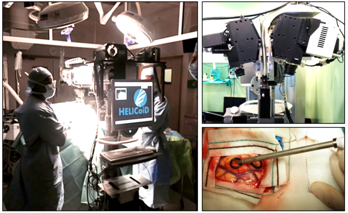
- Design of intraoperative visualization systems with HSI to highlight and delineate tumor margins in the operating room (Sensors 2018).
- Introduction of new algorithms for manifold embedding and semantic segmentation: segmenting hyperspectral data efficiently and ensuring consistency with real morphology (IEEE TMI 2017).
- Synthesis of the first clinical cohorts and real results from the HELICoiD project for introducing HSI in the operating room (IJS 2016).
Related Scientific Articles (with short description)
- Spatio-spectral classification of hyperspectral images for brain cancer detection during surgical operations
Introduces a supervised pipeline leveraging spatial and spectral features for automatic classification of tumor tissue. Improves intraoperative detection and surgical safety. - In-Vivo Hyperspectral Human Brain Image Database for Brain Cancer Detection
Describes creation and release of the first in-vivo HSI brain dataset from patients, published to foster the development and validation by the international community. - An intraoperative visualization system using hyperspectral imaging to aid in brain tumor delineation
Presents a real system for visualization and interpretation of HSI data at the operating table, integrating automatic analysis and clinical decision support. - Manifold embedding and semantic segmentation for intraoperative guidance with hyperspectral brain imaging
Introduces new strategies for data compression through manifold embedding and a robust semantic segmentation pipeline for real-time brain tissue segmentation. - Intra-operative hyperspectral imaging for brain tumour detection and delineation: Current progress on the HELICoiD project
First clinical summary of intraoperative HSI results collected in the European project HELICoiD, real cohort testing, and defining the roadmap for clinical technology integration.
Resource & Team
- Open-access HSI Human Brain dataset: IEEE Access 2019
- Code/tools available upon request from the authors.
- Daniele Ravì (segmentation, project design, supervision)
- H.Fabelo, G.M. Callicó, S.Ortega, G-Z Yang, D. Bulters, J.F. Piñeiro, R. Lazcano, E. Juárez, S. Kabwama, BR Kiran, C. Sosa, A. Szolna, etc.
Multidisciplinary collaboration and sharing of public datasets to accelerate international research into intelligent imaging and surgery.